
It’s official! Thanks to our people, we’re proud to have earned Great Place to Work® Certification™. Our company culture is our top priority! #GPTWcertified
")
About Berkshire Veterinary Hospital
Berkshire Veterinary Hospital is a leading-edge full-service veterinary facility offering 24-hour patient care and emergency services to our current clients. Utilizing the latest in veterinary technology, we compassionately deliver medical and surgical care in areas such as cardiology, dermatology, ophthalmology, internal medicine, dentistry, reproductive and transfusion medicine, and soft tissue and orthopedic surgery for dogs, cats, birds, and exotic pets. Our veterinary staff also offers consultation and training for dog and cat owners in the management of behavioral problems.
Complete Veterinary Care in Pittsfield, MA
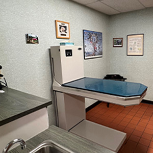
Berkshire Veterinary Hospital is proud to be on the leading edge of veterinary medical and preventative care. Our 6500 square foot facility located on five acres in Pittsfield, Massachusetts, is equipped with some of the latest in medical technology for your pet’s health.
Pet Wellness Exams
Preventative care is vital to the health of your pet. We recommend that all dogs and cats receive a physical exam.
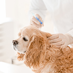
Pet Vaccinations
Vaccinations are important to help protect your pet and give them longer, healthier lives.
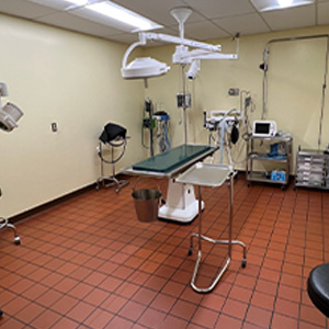
Pet Surgery
Berkshire’s staff is highly skilled and prepared for your pet’s surgical needs.
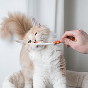
Pet Dental Care
Routine dental cleanings are vital in maintaining your pet’s oral health as well as its overall health.
Thank you for all your kind words.
Meet Our Veterinary Team
At Berkshire Veterinary Hospital, our veterinary team will invest the necessary time to provide the most skilled pet care by utilizing cutting-edge methods. We give the highest level of care for your pets with dependable abilities and kind touch, just like we would for our own pets.
")